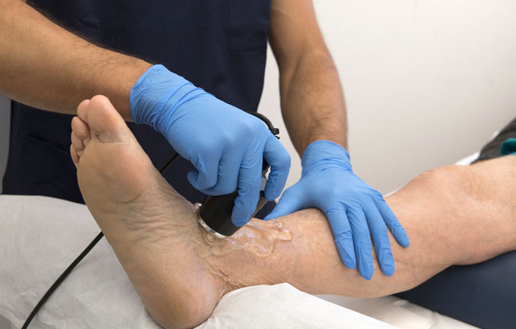
State-of-the-Art Ultrasound for Foot & Ankle
Doctors use a number of different imaging procedures to look at the structures within our feet and ankles. One of the most commonly used of these procedures is foot and ankle ultrasound which uses high-frequency sound waves to form an image of the bones and other parts of the foot and ankle. Ultrasound works by focusing these high-frequency sound waves on the area the doctor wishes to examine. The sound waves bounce off the bones and soft tissues and are picked up by the same device that emitted them; this device is called a transducer. The transducer sends this information to a computer, which uses it to form an image.

Although it can be used to diagnose fractures and other conditions related to bones, ultrasound is more commonly used to look at problems with soft tissue. Foot and ankle ultrasound examination can be useful for diagnosing and treating the following conditions, among others Morton’s neuroma, tendonitis, bursitis, heel spurs, tarsal tunnel syndrome, ankle injuries involving ligaments or tendons, and plantar fasciitis.
Ultrasound-Guided Injections
Ultrasound is not only useful in the examination and diagnosing of conditions but also in aiding the treatment. Ultrasound is one of the most commonly used devices for image-guided injections. Physicians often use injections to treat a variety of conditions. Commonly injected medications include cortisone and local anesthetics. Joint injections, for example, need to be administered within the joint space and not the surrounding soft tissue. Similarly, tendon injections should be administered in the tendon sheath, the structure covering the tendon, and not the tendon itself. The ultrasound also allows you to visualize fluids so that you can see if the medication is being distributed exactly where you need it to be.


